
BCC-NAC is more common on left breast than right ( 5). The male: Female ratio is 1:1 in BCC for common sites but BCC-NAC shows male preponderance which is thought to be because of increased exposure of chest area to sun light ( 1, 3- 7). ( 1, 7).īCC-NAC in males is commonly seen between 43-80 years of age and in females between 49-75 years of age. From 1983 to 2011 40 cases are reported in English literature of which 25 cases are males with one third of cases were solely with nipple involvement. Robinson reported first case of a rodent ulcer of the male breast in a 60 year old male in 1893 ( 3, 8). Basal cell carcinoma of nipple areola complex (BCC-NAC) is extremely rare and increasing ( 1- 5, 7).
#Itching mole on breast skin
Patient was followed up for one year, no recurrence or metastasis noted.īCC is the common malignancy of skin especially in western world and the incidence continues to increase worldwide ( 1- 3, 5). A final histopathological diagnosis of Pigmented Basal cell carcinoma of nipple and areola was made. (Fig.4) 4) and positive for pankeratin. Immunohistochemistry of tissue sections were negative for HMB45 (Fig.
#Itching mole on breast free
The resected surgical margins were free from the tumour. Foreign body giant cell reaction was seen. Stroma showed infiltration by chronic inflammatory cells and melanophages (Fig. Focal areas showed melanin pigment in tumour cells. The tumour cells were also arranged as interconnecting cords in between myxoid matrix. The tumour cells were round to oval with vesicular nuclei and scanty cytoplasm with peripheral pallisading arrangement. Epidermis showed proliferating basaloid cells, extending into dermis. Tissue sections were taken from paraffin blocks, which were dewaxed by xylene, dehydrated with isopropyl alcohol and stained with hematoxylin and eosin stain by routine conventional method. The tissue bits were processed by routine processing method using increasing grades isopropyl alcohol as dehydrating agent, xylene as clearing agent, embedded in paraffin wax and tissue blocks were prepared with paraffin wax. Cut section over pigmented lesion showed grey white to grey black areas. Pigmented lesion over nipple and areola measured 1 × 0.5 cms and showed ulceration.
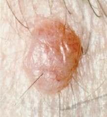
:max_bytes(150000):strip_icc()/molelightA500-56a2453a5f9b58b7d0c878e9.jpg)
Gross specimen consisted of elliptical skin with subcutis measuring 7 × 3.5 × 2 cms with nipple and areola. The specimen was subjected to histopathological examination. Patient underwent surgical three dimensional excision along with nipple and areola with one cm margin and primary closure. Mammography and ultrasound findings were not contributory. A provisional clinical diagnosis of melanoma was made. No past history of burns, surgery, trauma, disease of nipple or skin cancer. No past history of exposure to radiation, arsenic or tar. No similar lesion found elsewhere in the body. Local examination showed pigmented macular lesion involving nipple and areola of left breast. This does not definitely mean you have cancer.A 78 year old male, RAN, from Bangalore, India, presented with a pigmented lesion over left chest wall associated with itching and burning sensation since 4 months, gradually increasing in size. This may be an urgent referral, usually within 2 weeks, if you have certain symptoms. The GP may refer you to a specialist in hospital for more tests if they think you have a condition that needs to be investigated.

They may ask you if they can take a photograph of it to send to a specialist (dermatologist) to look at. The GP will look at your mole and any other areas of affected skin. Also tell them if you or a member of your family have had skin cancer in the past. Tell the GP if you have a mole, freckle or other area of skin that's recently changed.

You will be asked some questions about your health, family medical history, medical conditions and your symptoms.


 0 kommentar(er)
